- Products
- Oncohematology
- Antibodies
- Kits
- CAR T-cell
- Euroflow
- Single reagents
- Request info
- Resources and support
- Immunology
- Antibodies
- Single reagents
- Cross match determination (FCXM)
- FcεR1
- Ig subclasses
- Single reagents
- Kits
- TiMas, assessment of tissue macrophages
- Request info
- Resources and support
- Antibodies
- Exosomes
- Accesory reagents
- Software
- Oncohematology
- Services
- Peptide Production
- Design
- Modification
- Protein Services
- Expression and purification
- Freeze drying
- Monoclonal And Polyclonal Antibody Development
- Monoclonal
- Policlonal
- Specialized antibody services
- OEM/Bulk production
- Purification
- Conjugation
- Custom Exosome Services
- Isolation and purification
- Characterization
- Peptide Production
- Shop
- Support
- About Us
- Contact
ExoStep Platform: Detection and Characterization of Exosomes
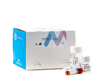
ExoStepTM KIT: The most accurate method of exosome detection developed to date.
ExoStepTM kit is the most accurate method of exosome isolation developed to date, a superior alternative for the sensitive detection of exosomes compared with the most commonly used methods. Our kit is easy to implement and analyse for any laboratory that has access to a conventional flow cytometer.
Specific Exosome Detection in biological fluids by Flow Cytometry
Wide dynamic range and limit of detection
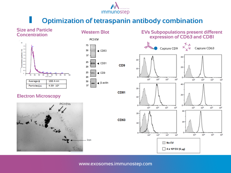
Allowing simultaneous Immunophenotyping of captured exosomes population
Easy to perform Immunobead assay with quantitative and scalable results
Specific and unambiguous exosome detection in biological fluids by flow cytometry:
ExoStepTM kit is an Immunobead assay for the detection of exosomes using a bead-bound capture antibody and fluorochrome conjugated detection antibody. This kit provides reproducible results and is intended for Flow Cytometry analysis of pre-enriched human exosomes from biofluids (plasma, serum, urine, among others) or cell culture media.
Ultracentrifugation is not needed
Reproducible results
Effective with small sample quantities
We are looking forward to hearing about your research
Bead-based assay for Flow Cytometry
ExoStepTM Kit for the Detection and Characterization of exosomes by Flow Cytometry is composed of an exosome capture reagent consisting of 6 µm diameter magnetic beads coated with a specific antibody as solid suport detectable by the cytometer. Additionally, the capture beads are dyed red and fluorescently labelled which facilitate their detection in the fluorescent detector of a conventional flow cytometer. ExoStepTM contains a detector antibody fluorescent conjugated for exosome detection.


Wide dynamic range and limit of detection
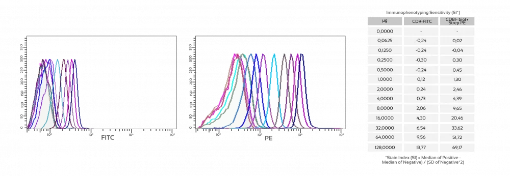
To determine the dynamic range and limit of detection of the assay, EVs titration experiments were performed using CD63 for capture and CD9 for detection. The sensitivity of the assay has demonstrated to be very high with a positive signal detected as little as thirty ng of exosomes while 2 ug were required for Western Blot detection. Besides, Flow Cytometry data follows a linear distribution between 30 and 8 micrograms of exosomes, allowing interpolating fluorescent intensity values to estimate the amount of exosomes in the sample.

Video Protocol
UNDER LICENCE FROM THE SPANISH NATIONAL RESEARCH COUNCIL (CSIC)
We work to apply our technology in the detection and quantification of disease biomarkers present in the surface of EVs as well as in diagnostic of diseases.
*This Technology is under patent licence: PET/EP2021/068633 “Method for the detection and/or quantification of extracellular vesicles in fluid biological samples”
Simultaneous Immunophenotyping
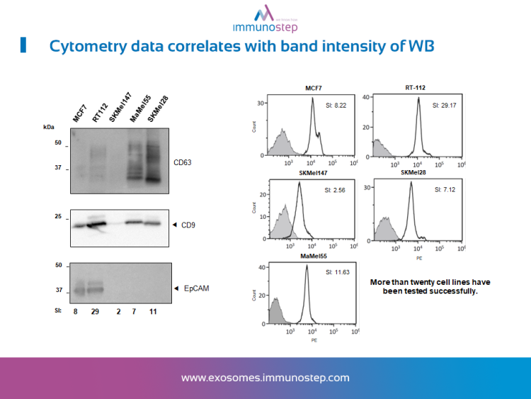
Considering exosome direct detection results in body fluids, we want to anticipate researcher’s needs for exosome subpopulations characterization through specific markers. In order to optimize the protocol, we performed an Immunophenotyping assay simultaneously with the exosome detection. This optimization of the protocol made possible to include in the FL2 channel (the most sensitive), the antibody against the marker of interest. The suitability of the kit for the specific detection of exosomes was confirmed, making possible to further characterize their phenotype by labelling with other markers of interest.
ExoELISA-Step Kit
ExoELISA-Step is an enzyme immunoassay for the detection and quantification of exosomes, based on the use of antigens or antibodies labeled with an enzyme, so that the resulting conjugates have both immunological and enzymatic activity. As one of the components (antigen or antibody) is labeled with an enzyme and insolubilized on a support (immunosorbent), the antigen-antibody reaction will remain immobilized and therefore, is easily revealed by the addition of a specific substrate. When acting on the enzyme a simple or quantifiable view will produce an observable color using a spectrophotometer or a colorimeter.

You might be also interested in:
References
| Kit Name | Kit Description | +info |
|---|---|---|
ExoELISA-Step Cell Culture | 96 test | EXO2506 | Cell culture Samples | ELISA Assays for detection and quantification of exosomes | | Go to shop |
ExoELISA-Step Serum/Plasma | 96 test | Exo2508 | Serum/plasma Samples | ELISA Assays for detection and quantification of exosomes | | Go to shop |
ExoStep Culture | 25 test | ExoS-25-C9 | Cell culture Samples | Superparamagnetic Capture Beads (CD63), Primary detection antibody (CD9 PE) and Assay Buffer 10X | | Go to shop |
ExoStep Serum/Plasma | Go to shop | |
ExoStep Urine | Go to shop | |
ExoStep Culture + Standard | 50 test | ExoS-50-CST9 | Cell culture Samples | Superparamagnetic Capture Beads (CD63 Capture Beads) Primary detection antibody (CD9 PE) Assay Buffer 10X Lyophilized exosomes (1×10^12) from PC-3 Human prostate cancer | | Go to shop |
ExoStep Serum/Plasma + Standard | Go to shop | |
ExoStep General Kit | 50 test ● 25 test | ExoS-50-G9 ● ExoS-50-G81 ● ExoS-25-G9 ● ExoS-25-G81 | Any biological Sample | Superparamagnetic Capture Beads (CD63 Capture Beads) Primary detection antibody (CD9 Biotin) or (CD81 Biotin) Secondary detection reagent (PE Conjugated) Assay Buffer 10X | | Go to shop |
Mouse ExoStep Culture | 25 test | MO2ExoS-25-C9 | RUO mouse exosome from Cell Culture Samples | Superparamagnetic Capture Beads (CD63 Capture Beads) Primary detection antibody (CD9 Biotin) Secondary detection reagent (PE Conjugated) Assay Buffer 10X | | Go to shop |
