- Products
- Oncohematology
- Antibodies
- Kits
- CAR T-cell
- Euroflow
- Single reagents
- Request info
- Resources and support
- Immunology
- Antibodies
- Single reagents
- Cross match determination (FCXM)
- FcεR1
- Ig subclasses
- Single reagents
- Kits
- TiMas, assessment of tissue macrophages
- Request info
- Resources and support
- Antibodies
- Exosomes
- Accesory reagents
- Software
- Oncohematology
- Services
- Peptide Production
- Design
- Modification
- Protein Services
- Expression and purification
- Freeze drying
- Monoclonal And Polyclonal Antibody Development
- Monoclonal
- Policlonal
- Specialized antibody services
- OEM/Bulk production
- Purification
- Conjugation
- Custom Exosome Services
- Isolation and purification
- Characterization
- Peptide Production
- Shop
- Support
- About Us
- Contact
-
Highly sensitive assays to detect early to late apoptotic stages;
-
Large number of assays to detect different parameters;
-
Very well validated in Flow Cytometry with many different type of cells;
-
Validated with many different types of drugs;
-
Different fluorescent options for multiple laser excitation sources.
Apoptotic cell detection: Diagnosis Application
Apoptosis is a regulated process of cell death that occurs during embryonic development as well as maintenance of tissue homeostasis. The appearance of non-regulated apoptosis involves different diseases, such as neurodegenerative diseases and cancer.

Neurological Diseases

Cancer Research

Sperm Viability

Drugs Development
Understanding the mechanisms of programmed cell death (also known as apoptosis) is an important aspect in the research of e.g., tissue homeostasis, tumour diseases, or the toxicity and efficacy of drugs, among others.
Why is Annexin V Staining used to detect apoptosis?
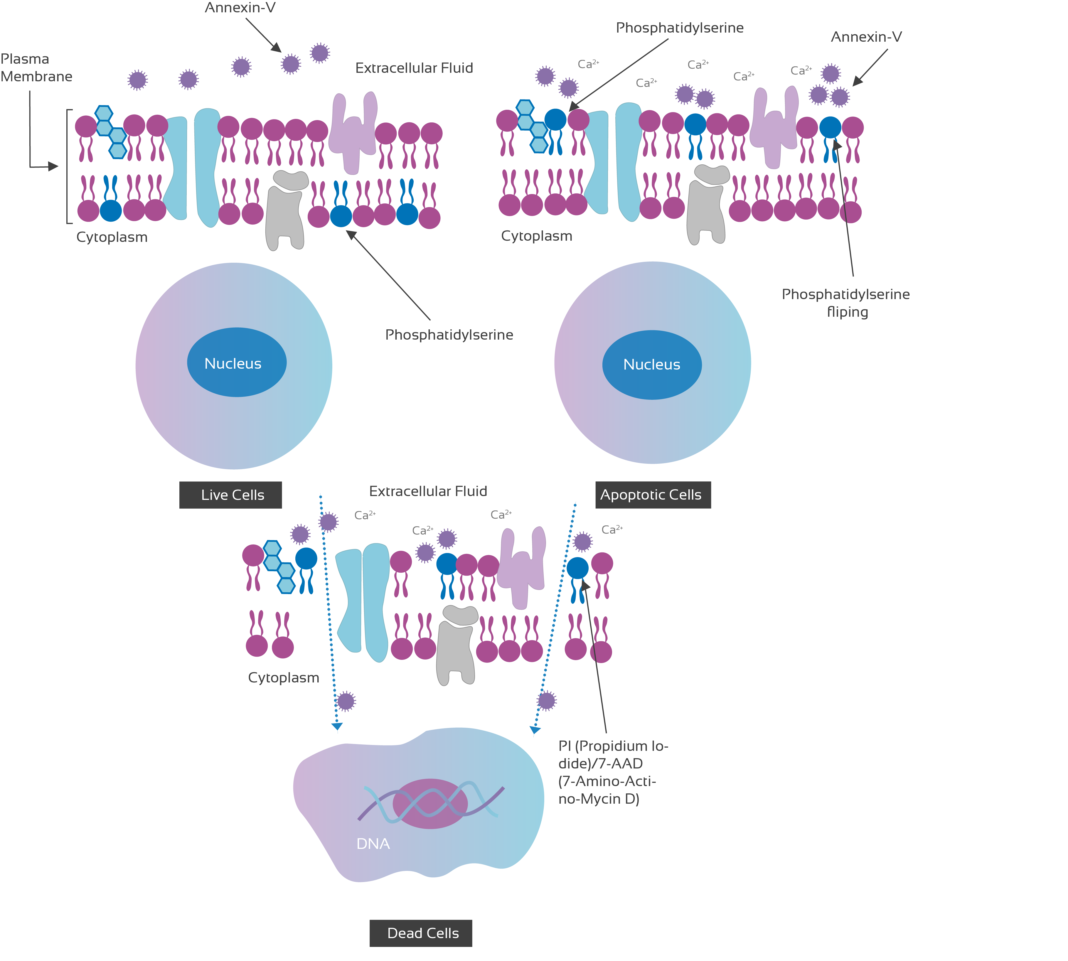
The human vascular anticoagulant, Annexin V is a 35-36KDa Ca2+ dependent phospholipids binding protein that has a high affinity for PS. In healthy cells, PS is located on the cytoplasmic surface of the plasma membrane.
However, during early apoptosis, phosphatidylserine (PS), normally located on the cytoplasmic surface of the cell membrane, becomes exposed to the extracellular environment, causing changes in the membrane asymmetry and permeability.

Highly sensitive assays to detect early to late apoptotic stages
The membrane assays from Immunostep are based on the combined detection of apoptosis and necrosis by fluorescent vital double staining with Annexin V and PI or 7-AAD. Annexin V binds to phosphatidylserine which is translocated outside of the membrane during apoptosis, while PI or 7-AAD can only penetrate into necrotic cells.
Flow Cytometry-based apoptosis detection
The flow cytometric analysis of apoptosis is based on different detection principles. Due to the complexity of cell death cascades, the multi-parameter kits from Immunostep are particularly well suited to assess the apoptosis.

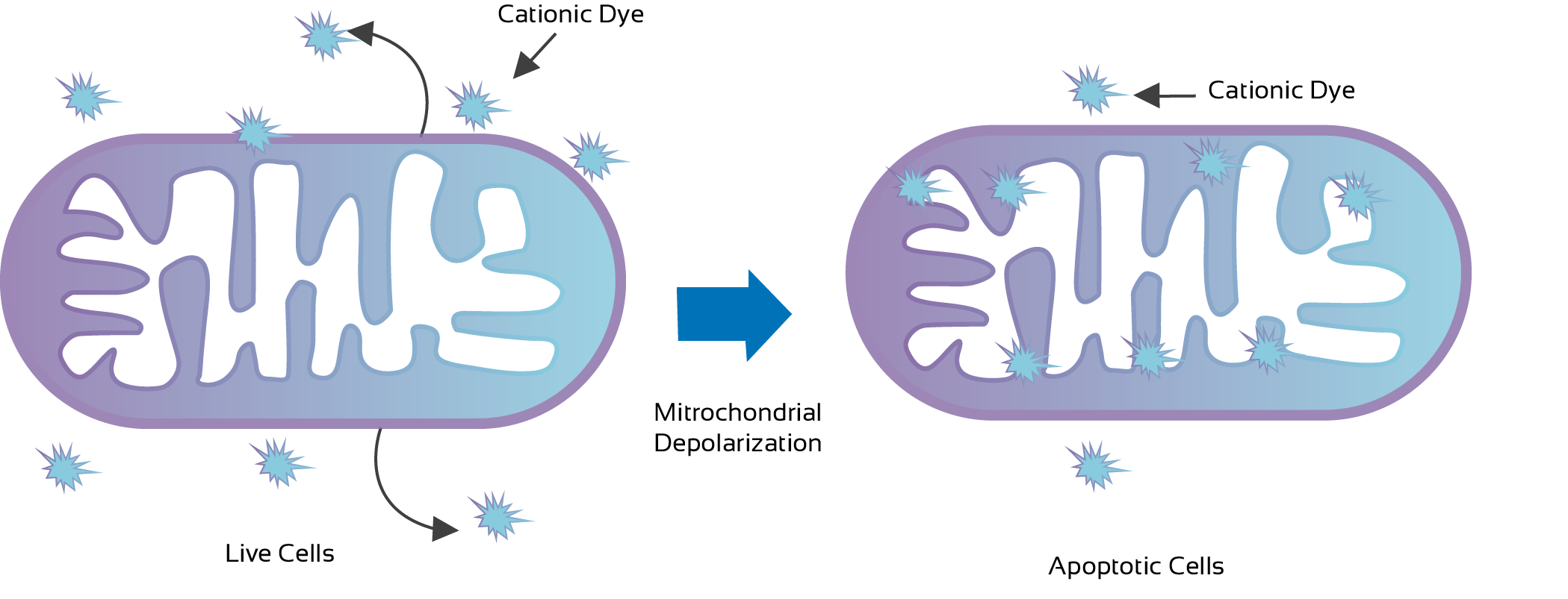
Figure 2: Mitochondrial cationic Dye process Illustration
Mitochondrial Assays: detection of early apoptotic cells
The dysfunction of the mitochondria is a special feature of earlier stages of apoptosis. MitoStep apoptosis kits measure changes in the mitochondrial membrane potential with a solution of the cationic cyanine dye DiIC1(5).
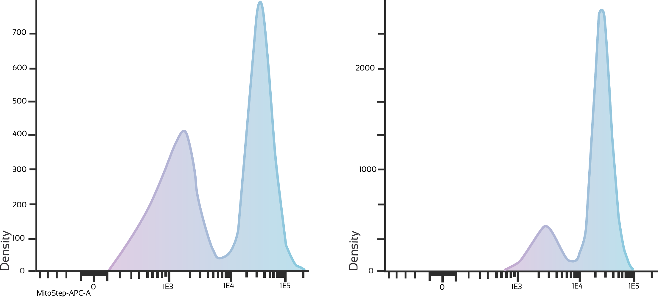
In healthy cells, especially in mitochondria with active membrane potentials, the dye accumulates. During apoptosis, the DiIC1(5) fluorescence intensity decreases and thus there is an increase of cells with lower DiIC1(5) fluorescence.
MitoStep Kits:
Depolarization of the submitochondria and thus changes in the mitochondrial membrane during the early stages of apoptosis have been described. This is the reason why mitochondrial uptake of cationic dyes is a possible source of fluorescence variation.
DiIC1(5)-stained cells can be visualized by excitation with a 633 nm laser and emission at 658 nm, and combined with FITC or PE staining.

Figure 3: Jurkat Cells T/cell Leukemia, human (treated with 6um Camptothecin for four hours/both panel.

PRODUCT REFERENCES
ANXVKF-100T | FITC Apoptosis Detection Kit | Download TDS | Go to shop |
ANXVKPE-100T | PE Apoptosis Detection Kit | Download TDS | Go to shop |
ANXVKDY-100T | Dy634 Apoptosis Detection Kit| Download TDS | Go to shop |
ANXVKCFB-100T | CF-Blue Apoptosis Detection Kit | Download TDS | Go to shop |
ANXVKCFB7-100T | CFBlue Annexin V Apoptosis Detection Kit with 7-AAD | Go to shop |
ANXVKF7-100T | FITC Annexin V Apoptosis Detection Kit with 7-AAD | Go to shop |
MITO-100T | MitoStep | Download TDS | Go to shop |
KMAF-100T | FITC MitoStep + Apoptosis Detection Kit | Download TDS | Go to shop |
KMAPE-100T | PE MitoStep + Apoptosis Detection Kit | Download TDS | Go to shop |
ANXVB-200T | Biotin Annexin V | Download TDS | Go to shop |
ANXVF-200T | FITC Annexin V | Download TDS | Go to shop |
ANXVKPE-200T | PE Annexin V | Download TDS | Go to shop |
ANXVKDY-200T | DY634 Annexin V | Download TDS | Go to shop |
ANXVKCFB-200T | CFBlue Annexin V | Download TDS | Go to shop |
BB10X-50ML | Annexin V Binding Buffer (10X) | Download TDS | Go to shop |


