What is an exosome?
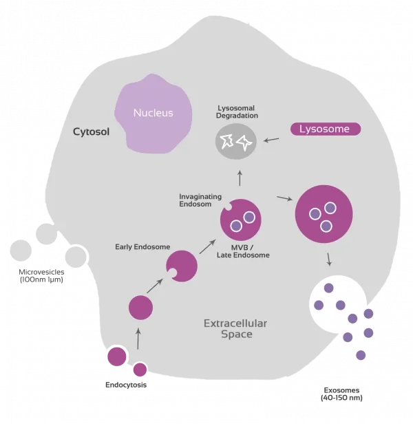
Exosomes are small (~40-150 nm) extracellular vesicles (EVs) released from all cell types and found in body fluids and cell culture supernatants. They are generated by fusion of a specialized endosome, the multivesicular body (MVB), with the plasma membrane.
Exosomes have been proposed to provide means for intercellular exchange of macromolecules, allowing the transfer of proteins, lipids, mRNA and miRNA, contributing to intercellular communication in relevant biological processes, including apoptosis, antigen presentation, angiogenesis, inflammation, and coagulation.
Exosome Research
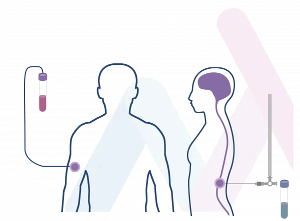
Exosomes have recently emerged as a new source of potential non-invasive biomarkers of several diseases, since they can be easily obtained from body fluids such as urine, blood, saliva, or breastmilk and their composition may be directly dependent on the physiological and/or pathological state of the patient.

Cellular Therapy

Active Ingredient in cosmetics

Targeted drug delivery

Regenerative Medicine

Disease monitoring: Liquid Biopsy

Biomarkers for several diseases
The potential of Exosomes in Cancer Research
A biomarker with high specificity and sensitivity, is a basic requirement for non-invasive cancer diagnosis. Exosomes are a type of lipid bilayer extracellular vesicles (EVs), containing different components, including proteins, lipids, DNA, messenger RNA (mRNA), and non-coding RNAs. Increasing evidence indicates that nucleic acids are protected by exosome lipid membrane. These vesicles are almost released from all cell types, into biological fluids.
Exploring the incredible growth potential of EVs as minimally invasive liquid biopsies
Exosome characterization and analysis present a tremendous clinical opportunity, specifically in the area of liquid biopsy. But this potential requires detecting and characterizing exosomes with high accuracy and reproducibility.
Immunostep makes available to its clients all the experience and technical capabilities in the field of exosomes to make the clinical use of these EVs a reality.


In 2019 Immunostep R&D team recognized the great potential of exosomes and since then we have been working on the development of the best methods for detection, quantification, isolation, characterisation and immunophenotyping of extracellular vesicles (EVs), that are present in biological fluid samples, using polycationic polymers and a solid surface functionalized with an EVs capturing molecule.
During the following years we have been working to apply this and other methods in the exosome research in order to participate in the improvement of the performance of these tools for different areas of research and diagnosis. Backed by our extensive expertise in exosomes, as an exosome reagent supplier worldwide we are still working on amplify our technology and transforming them into powerful tools that contribute and help exosome research around the world.