- Products
- Oncohematology
- Antibodies
- Kits
- CAR T-cell
- Euroflow
- Single reagents
- Request info
- Resources and support
- Immunology
- Antibodies
- Single reagents
- Cross match determination (FCXM)
- FcεR1
- Ig subclasses
- Single reagents
- Kits
- TiMas, assessment of tissue macrophages
- Request info
- Resources and support
- Antibodies
- Exosomes
- Accesory reagents
- Software
- Oncohematology
- Services
- Peptide Production
- Design
- Modification
- Protein Services
- Expression and purification
- Freeze drying
- Monoclonal And Polyclonal Antibody Development
- Monoclonal
- Policlonal
- Specialized antibody services
- OEM/Bulk production
- Purification
- Conjugation
- Custom Exosome Services
- Isolation and purification
- Characterization
- Peptide Production
- Shop
- Support
- About Us
- Contact

Custom Exosome Services
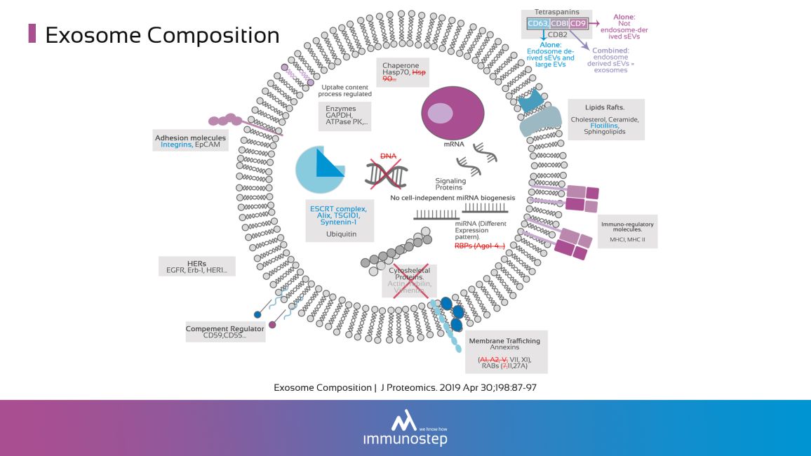
Exosomes are small (~40‐150 nm) extracellular vesicles (EVs) released from all cell types and found in body fluids and cell culture supernatants. Exosomes are generated by fusion of a specialized endosome, the multivesicular body (MVB), with the plasma membrane. Exosomes have been proposed to provide means for intercellular exchange of macromolecules, allowing the transfer cellular information, contributing to intercellular communication in relevant biological processes.
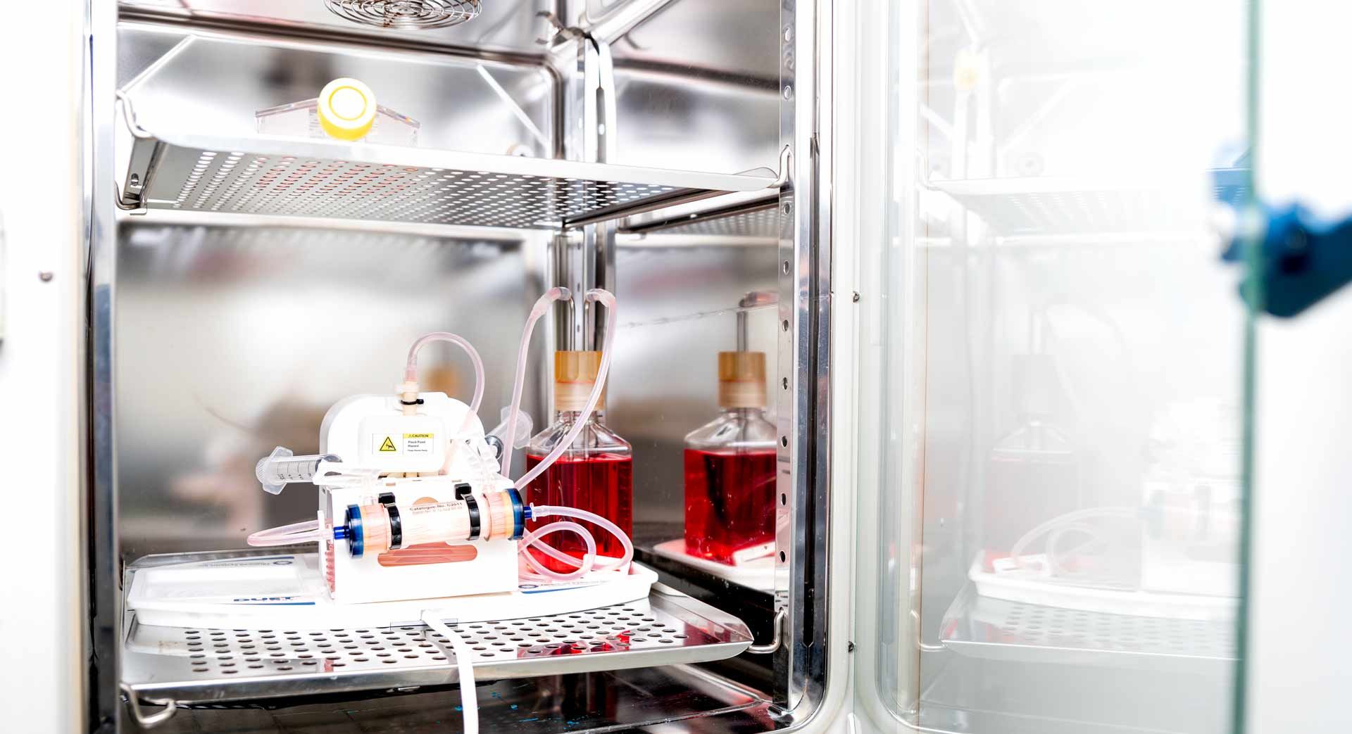
Exosome Isolation and Purification
Immunostep has developed a series of protocols for the isolation and purification of high-quality exosomes derived from multiple sources including cell culture samples and all human proximal fluids, including plasma, urine, cerebrospinal fluid, amniotic fluid, bronchoalveolar lavage fluid, synovial fluid, sperm, saliva, malignant and pleural effusions of ascites and breast milk.

Based on density and size under centrifugal force is generally considered being the gold standard method for extracellular vesicles isolation, although it may produce some aggregation of EVs.
Based on EVs solubility, is a quick method, with high recovery although it usually precipitates non-EV material.
This is probably the most suitable method, alone or combined, to isolate exosomes from biological samples and in particular from plasma/serum, since it allows the separation of exosomes from important contaminants such as proteins and HDL. Additionally, Immunostep has columns that allow separation between EVs-Exosomes and other Extracellular Vesicles.
Based on the use of capture beads, it allows the specific isolation of exosomes based on the membrane display of specific marker. For example: CD63, EpCAM, PD-L1, etc.
Protocols for Exosome Purification
At Immunostep we have developed a wide range of protocols for exosome isolation/purification from all kind of samples, providing a customized solution for each project, with high purity and optimal recovery results and compatible with any downstream processing or analysis.
WE ARE LOOKING FORWARD TO HEARING ABOUT YOUR EXOSOME RESEARCH
Characterization Methods
We have developed and implemented several characterization and validation methods, for both research and clinical purposes, that allow analyze purity and composition and to quantify exosomal cargo, which contains critical information that has proved to be tremendously promising for diagnostic, prognostic and therapeutic use in a variety of disease areas. These methods include:
-
Transmission electron microscopy (TEM)
-
Scanning electron microscopy (SEM)
-
Nanoparticle tracking analysis (NTA)
-
Western blot (WB)
-
Flow cytometry
-
Fluorescence-activated cell-sorting (FAQS)
-
Multiplexing Bead Arrays
-
Enzyme-linked immunosorbent assay (ELISA)
-
Gene expression analysis (mRNA and microRNA)


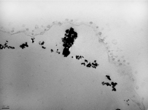
Electronic microscopy: exosomes isolated by a capture bead


Fluorescence: activated cell sorting (FACS)


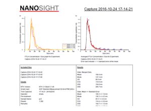
Nanoparticle tracking analysis (NTA).Size and particle concentration analysis report.
Exosome Characterization
Exosome characterization and analysis presents a tremendous clinical opportunity and specifically in the area of liquid biopsy. However, realizing the potential requires detecting and characterizing exosomes with high accuracy and reproducibility, which can be challenging for any project or research. Immunostep makes available to its clients all their experience and technical capabilities in the field of exosomes, to make the clinical use of these EVs a reality.
