The role of exosomes in cancer – Part 2
Let’s continue where we left off in our last article. We were talking about exosomes in liquid biopsies, and its relation with cancer.
In oncology, one of the biggest challenges is to get an early diagnosis, but not only, as it is also desired to avoid side effects of invasive biopsies, the excess of treatments, and psychological stress of patients.
▌Exosomes and liquid biopsy
Bearing in mind all the aspects, the exosomes have a great potential to serve as a liquid biopsy tool in these diseases. In particular, tumoral exosomes most likely are useful as biomarkers for early cancer detection, after all they carry the content that reflects genetic or signaling alterations in the cancer cells in which they are originated from.
Exosome-based liquid biopsy deserves consideration on conventional tissue biopsy for various reasons, as it is non-invasive in contrast to tissue biopsy, which requires invasive surgery and only provides a limited amount of a local area of the tumoral tissue that does not reflect the tumor heterogeneity. Besides, exosomes are easily to detect and obtained from bodily fluids (ie. pheripheral blood, …) and tumor exosomes provide dynamic information of the tumor heterogenecity at the time of extraction. Furthermore, due to exosome stability in body fluids, exosome-based diagnosis provides greater sensitivity than other approaches.
▌Exosomes in tumoral pathologies
It has been already demonstrated in highly relevant solid tumors, such as Prostate Cancer, Colorectal Cancer, Lung Cancer, Melanoma, Ovarian Cancer or Breast Cancer, which could be helpful to establish an early diagnostics or disease monitoring.
PROSTATE CANCER
For example, Prostate Cancers (PCa) present a highly heterogeneous phenotype, so that the genotype in particular area of the tumor may be high different from other areas. PSA test is the gold standard for the early detection of PCa, however it has led to a considerable number of over diagnoses and excessive treatments, since PSA levels are often elevated in patients with benign or non-cancerous conditions, such as benign prostatic hyperplasia, infections or chronic inflammation. On this matter, the content of circulating PCa tumor exosomes in plasma and urine has been described, with specific markers, such as caveolin-1 (Cav-1), prostate-specific antigen (PSA), PCA3 and fusion protein TMPRSS2-ERG, or Survivin among others, together with exosomal markers such as CD81 or CD63, in PCa patients.
COLORECTAL CANCER
About colorectal cancer cells (CRC), the secreted EVs into the bloodstream, are characterized by the presence of phenotype markers such as CD147 and CD9, which would allow it specific detection. On the other hand, and as occurs in PCa, Tumor-specific antigens have also been detected in CRC tumor exosomes, such as carcinoembryonic antigen (CEA) that has been described in ascites exosomes. Also in pancreatic cancer patient’s serum, enriched tumor exosomes with identified protein markers such as the macrophage migration inhibiting factor factor (MIF) or glypican-1 (GPC1) have been determined. The latest highlighted biomarker is relevant for specific and sensitive distintion between patients with cancer in different stages and patients with another type of pancreatic disease.
Additionally, there is also another evidence that GPC1 is a pan-specific biomarker of tumor exosomes, which makes it a powerful tool for the early diagnosis of the disease, permitting detection and isolation of these circulating exosomes for a later evaluation of genetic or protein profiles alterations.
LUNG CANCER
To date, lung cancer (LC) accounts for more deaths each year than any other type of cancer and its 5-year survival rate is low, because most patients with non-small cell lung cancer (NSCLC) show an advanced stage of disease. Even so, diagnosing NSCLC is still challenging and new methods are needed to support the clinical outcome already used to ensure a better overall result. Recently, this field has been encouraged with the possibility of using tumor exosomes as diagnosis and prognosis disease markers. Specifically, a panel of 12 miRNAs and membrane proteins such as CD91, CD317 and EGFR has been suggested as possible NSCLC exosomal markers. Similarly, it has been described how lipopolysaccharide-binding proteins (LBP) on exosomes can distinguish, more precisely than circulating LBP, between patients with metastatic vs non-metastatic patients NSCLC.
Finally, tumor exosomes containing proteins involved in the immunosuppression of the immune system, such as the T-cell molecule immunoglobulin, mucin-domain containing-3 (Tim-3) and its ligand Galectin-9, have also been detected in the plasma of melanoma patients. In fact, an increament of exosomes number, together with high expression levels of Galectin-9, has been correlated with greater tumor malignancy, together with high levels of exosomes containing Tim-3, has been correlated with the presence of lymph node metastases.
MALIGNANT MELANOMA
Malignant melanoma (MM) is an aggressive cancer with an increasing incidence and a poor prognosis. The most frequently used prognostic markers are S100 calcium-binding protein B (S100B), Melanoma inhibitory activity (MIA) and lactate dehydrogenase (LDH) and although they are the most used tumor markers for prognosis and follow-up in advanced MM, they present serious specificity and sensitivity limitations. In this sense, the presence of S100B and MIA has been detected in the protein content of exosomes obtained from patients with MM. Additionally, it has been observed that the sensitivity and specificity for the diagnosis of MM, through the detection of tumor exosomes with the S100B or MIA content, is superior to the analysis of the concentration of these biomarkers in serum. Previously, other authors had shown increasing amounts of exosomes expressing constitutive proteins such as CD63 and Rab-5b, as well as to a marker associated with tumors such as Cav-1, in MM patient plasma, comparing with healthy controls. This suggests the possible use of exosomes as diagnostic or prognostic markers of the disease. Metastatic MM or stage IV MM is an intractable cancer that lacks of reliable and non-noninvasive markers of disease progression. As in other tumors, there is presence of exosomes in the plasma of these patients. So, it would be logical to think that derived tumor exosomes from MM cells could play a relevant role in the monitoring of metastatic disease. In this direction, important advances have been performed, such as the identification of a specific biofirm of circulating turmoral exosomes derived from advanced MM, consisting of TYRP2, VLA-4, HSP70, an HSP90 isoform and MET, which would allow analysis of tumor content, besides the fact that exosomes YRP2 and MET, plus a high amount of protein per exosome, would enable predicting the capacity of disease progression [32]. Recently, NKG2D Ligand detection in metastatic MM derived exosomes, specifically MHC class I chain-related protein A (MICA), has been described, opening the possibility to be used as an exosomal tumor associated marker with potential clinical value.
OVARIAN CANCER
The majority of women diagnosed with ovarian cancer (OvCa) already have an advanced disease, which limits treatment options and contributes to a high risk of OvCa mortality, so the detection of tumor exosomes could be a boost necessary in the early diagnosis of the disease. As in other tumors, exosomes have been shown to express ovarian tumor-associated markers such as CA-125, EpCAM, CD24 and besides it has been described that they express TGF-β1 and MAGE3/6. This molecular profile could serve as markers of the presence and progression of the disease.
BREAST CANCER
In the same way, Breast cancer is the most common among women. The prognosis of breast cancer is based on its initial stage at the time of diagnosis with the main associated prognostic factors being lymph node involvement, tumor size and histological grade. However, the tumor in the same stage may behave differently, and the forecast may vary in a different evolution. Therefore, it is important to find biomarkers that predict the likelihood of recurrence and identify those patients who might benefit from an additional therapy. In this sense, the analysis of tumor exosomes can be of vital importance, since their number has been observed to increase in these patients, expressing selective patterns with markers such as CD24, EpCAM, EDIL3, Fibronectin. Additionally, a differential expression of Survivin has even been observed in exosomes in particular Survivin-2B, at different stages of the disease may serve as a diagnostic and/or prognostic marker.
CENTRAL NERVOUS SYSTEM
Of particular importance is the evaluation of exosomes in tumors of the central nervous system, which lie behind a partially intact bloodbrain barrier, which often prevents the release of CTCs. However, high numbers of EVs have been found in the blood of patients with glioblastoma multiforme (GBM), which offers new hope for treatment control of this devastating disease. With this aim, it has been analyzed the protein profile of EV derived from GBM, including exosomes, to see if there were differences between EV protein profile and cancer cell origin and with non-pathological cells, leading to a molecular signature distinct from the EVs derived from GBM, which present high expressions of epidermal growth factor receptor (EGFR), platelet-derived growth factor receptor (PDGFRα), podoplanin (PDPN), ephrin type-A receptor 2 (EphA2), and specific expression of Epidermal Growth Factor Receptor Variant III (EGFRvIII) and cytosolic isocitrate dehydrogenase 1 mutation (IDH1 R132H).
MYELOID LEUKEMIA
As previously detailed, exosomes play an important role in the suppression of the tumor immune response. In the same way, it has been described that patients newly diagnosed with acute myeloid leukemia (AML) have elevated levels of plasma exosomes with a specific phenotype consisting of transforming growth factor beta 1 (TGFb-1), latency-associated protein (LAP), Mature TGFb-1, CD9, CD81, CD34, CD117 and GAPDH, among which stands out the high content of TGFb-1 that would have a suppressive function of the cytotoxic activity of NK cells. Specifically, TGF-b1 expression levels in tumor exosomes correlate with the response to chemotherapeutic treatment and could eventually help to identify the presence of minimal residual disease in patients in complete remission.
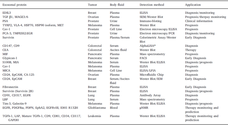
Finally, in addition to proteins (Table 1 below), analysis of protein/lipid proportions in exosomes can be used to further isolatation and characterization of different exosomes subpopulations present in body fluids. Similarly, there are studies characterizing the glycan composition of EV, and changes in this composition could be indicators of disease status, and could be used as diagnostic markers.
▌Identification of biomarkers
Phenotyping of exosomes is particularly important in the determination of cellular and subcellular origin, providing clues about the characterization of the different exosomal subpopulations, among them the tumor ones, as well as their functionality and the identification of biomarkers related to the disease. And this is the topic we will be talking about on our next article, stay tuned!
► Immunostep products related with this article:
- ExoStep Platform: https://immunostep.com/exosomes/exostep-platform-detection-and-characterization/
- Assay Custom products: https://immunostep.com/custom-services/custom-exosome-services/
- Exosome Isolation Columns: https://immunostep.com/exosomes/exosome-isolation-columns-sec/
► References:
- Jara-Acevedo, R., Campos-Silva, C., Valés-Gómez, M., Yáñez-Mó, M., Suárez, H., & Fuentes, M. (2019). Exosome beads array for multiplexed phenotyping in cancer. Journal of proteomics, 198, 87–97. https://doi.org/10.1016/j.jprot.2018.12.023